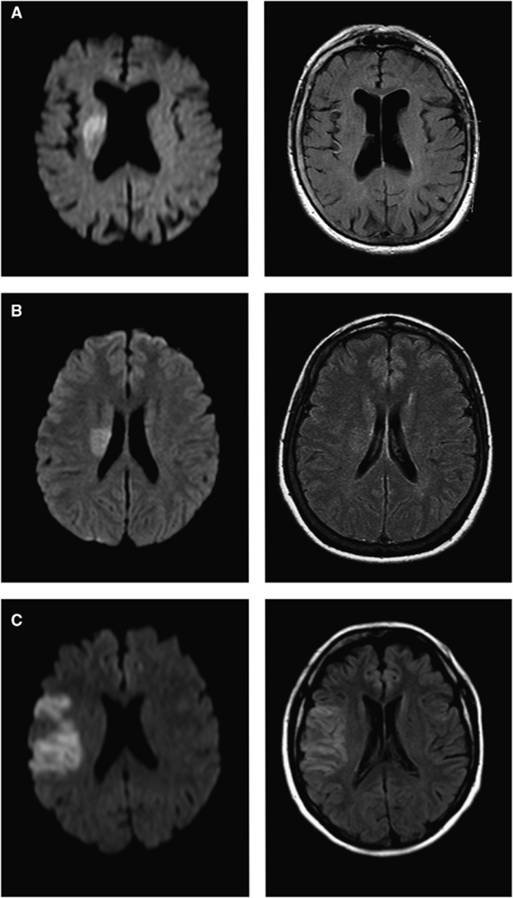
Figure 1.
Examples of diffusion-weighted imaging (DWI) and fluid-attenuated inversion recovery (FLAIR) images. DWI (left column) and FLAIR (right column) images. Acute ischemic lesions are easily identified on all DWI. FLAIR images show (A) no corresponding FLAIR lesions; (B) ‘subtle' FLAIR lesions; and (C) ‘obvious' FLAIR lesions.

