Figure 5.
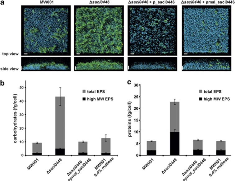
EPS analysis of the S. acidocaldarius deletion strain saci0446. (a) Three-day-old biofilms of trans-complemented Δsaci0446+p_saci0446 and Δsaci0446+pmal_saci0446 strains were analysed by CLSM and compared with the reference strain MW001 and Δsaci0446 strains. Biofilm cells were stained using 4′-6-diamidino-2-phenylindole (blue), ConA (green) and IB4 (yellow). The overlay images of all three channels are shown. Scale bar=20 μm. (b) EPS were isolated from 3-day-old biofilms of MW001, Δsaci0446, Δsaci0446+pmal_saci0446 and MW001 suplemented with 0.4% (w/v) of maltose. Carbohydrate concentrations were determined from both, non-dialyzed EPS (total EPS, gray bars) and dialyzed EPS extracts (3.5 kDa; high-molecular-weight EPS, black bars). (c) Protein concentrations of the same non-dialyzed and dialyzed EPS.The means and s.d. of three biological replicates are shown for b and c.

