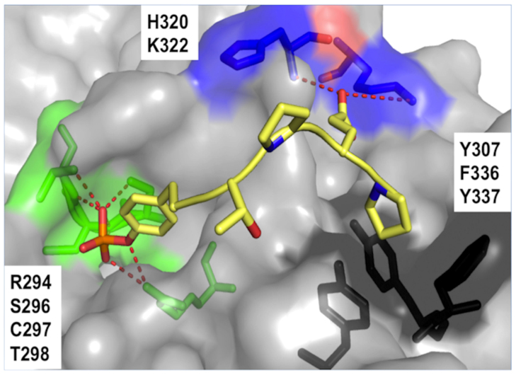
Figure 1. Structure of the complex between Cbl(TKB) (gray surface) and the pentapeptide (yellow sticks, PDB ID 4GPL).
The interacting residues at the Cbl(TKB):peptide interface are labeled and are represented as green sticks for the pTyr site, blue sticks for the p+3 Glu site, and black sticks for the p+4 Pro site. H-bonds are shown as red dashes.

