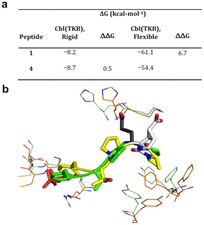
Figure 5. Computational studies of peptides 1 and 4 binding to Cbl(TKB) using MD and docking simulations.
(a) Comparison of 1 and 4 on of the change in binding energy when receptor is rigid or flexible. (b) Bound conformation as determined by MD. Green and orange lines indicate rigid and flexible Cbl(TKB), respectively. Green and yellow sticks represent 1 and 4, respectively. The variable amino acid at the p+3 position is shown as black sticks for peptide 1 and white sticks for peptide 4.

