
Figure 5. Degradation of Aβ40 within endosomal/lysosomal pathaway of bEnd3 and hCMEC/D3 cells.
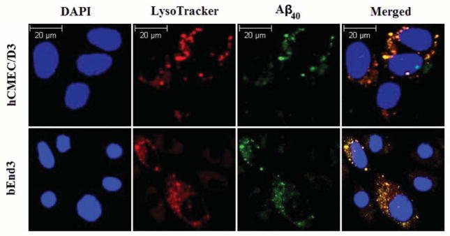
Co-localization of intracellular Aβ40-HiLyte Fluor and LysoTracker Red in endosomal/lysosomal pathway of brain endothelial cells (bEnd3 and hCMEC/D3) analyzed using confocal immunoFluorescence microscope. bEnd3 cells and hCMEC/D3 were incubated with 1 nM of Aβ40-HiLyte Fluor for 15 min or 12 h; respectively. DAPI stains nuclei. Lysosomes were labeled with LysoTracker Red for 45 min before the end of study. The yellow color in the merged image represent co-localization of green (Aβ40-HiLyte Fluor) and red (LysoTracker Red) fluorescence.