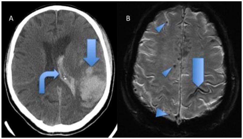
Figure 1.

Hemorrhagic findings of 2 patients with pathologically proven cerebral amyloid angiopathy. A, Head CT showing acute lobar parenchymal macrobleed (arrow) and intraventricular hemorrhage (bent arrow). B, GRE MRI shows lobar microbleeds (arrowheads) and sulcal siderosis (pentagon).
