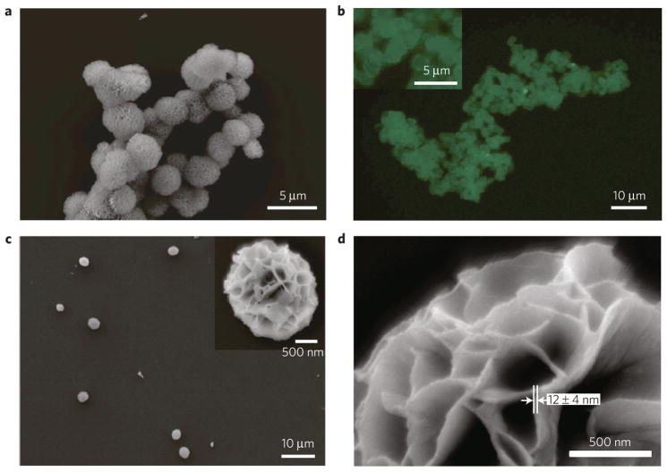
Figure 2. Characterization of the RNAi-microsponge.
a, SEM image of RNAi-microsponges. b, Fluorescence microscope image of RNAi-microsponges after staining with SYBR II, an RNA-specific dye. c, d, SEM images of RNAi-microsponges after sonication. Low-magnification image of an RNAi-microsponges (c). High-magnification image of an RNAi-microsponge (d).

