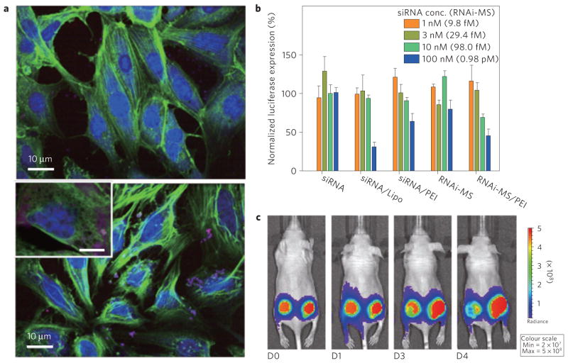
Figure 5. Transfection and gene silencing effect.
a, Intracellular uptake of red fluorescent dye-labelled RNAi-microsponges without PEI (top) and RNAi-microsponge/PEI (bottom). To confirm the cellular transfection of RNA particles, both types of particles, labelled for red fluorescence, were incubated with T22 cells. Fluorescence labelled RNAi-microsponges without a PEI outer layer showed relatively less cellular uptake by the cancer cell line (T22 cells) suggesting that the larger size and strong net negative surface charge of RNAi-microsponges probably prevents cellular internalization. Inset: scale bar, 5 μm. b, Suppression of luciferase expression by siRNA, Lipofectamine complexed with siRNA (siRNA/Lipo), siRNA complex with PEI (siRNA/PEI), RNAi-microsponge (RNAi-MS), and RNAi-microsponge condensed by PEI (RNAi-MS/PEI). The same amount of siRNA is theoretically produced from RNAi-microsponges at the concentration in parentheses. c, In vivo knockdown of firefly luciferase by RNAi-MS/PEI. Optical images of tumours after intratumoral injection of RNAi-MS/PEI into the left tumour of a mouse and PEI solution only as a control into the right tumour of the same mouse.

