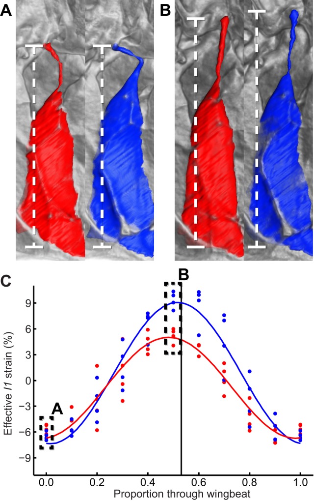
Figure 9. Buckling of the I1 tendon.

(A, B) Visualizations of the I1 muscle at the start and end of the downstroke, respectively. The tendon is buckled on both wings at the start of the downstroke (A) but has been pulled straight by the end of the downstroke (B). Each panel compares the state of I1 on the high-amplitude (blue) and low-amplitude (red) wing. (C) effective strain measured along the straight line joining the attachment points of I1 (dashed line). Comparing the amplitude of this effective strain with the amplitude of the actual I1 muscle strain in Figure 6F shows that the buckling tendon accommodates a 4-fold enhancement in the range of movement of the first axillary sclerite on the high-amplitude wing.
