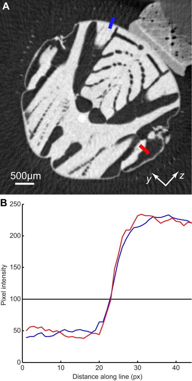
Figure 10. Edge detail of two parts of the thorax.

(A) Tomogram showing transverse section of the thorax, with the mount visible in the upper right of the image. The blue line cuts through the scutum, which is a rigid part of the thorax that did not move measurably during recordings. The red line cuts through the steering muscles, which oscillate at wingbeat frequency. (B) Pixel intensities along the two lines indicated in (A). Edge sharpness, as measured by the steepness of the change in pixel intensity along each line, is essentially identical for the scutum and the steering muscles. This indicates that the position of the steering muscles must have been consistent between wingbeats, at the phase of the wingbeat shown here, which indicates that the steering muscle kinematics did not vary measurably between wingbeats.
