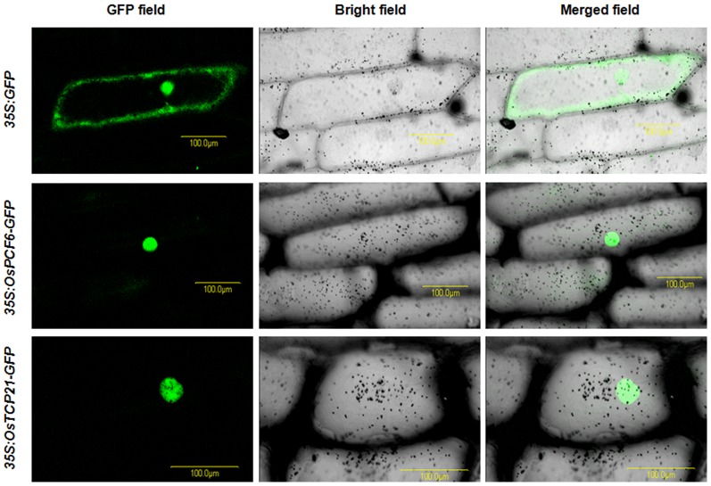
Figure 10. OsPCF6-GFP and OsTCP21-GFP proteins were located in the nuclei.
Confocal images of onion epidermis cells under the GFP channel showing the constitutive localization of GFP and the nuclear localization of OsPCF6- and OsTCP21-GFP fusions driven by the cauliflower mosaic virus 35S promoter. Onion epidermal peels were bombarded with DNA-coated gold particles and GFP expression was visualized 24 h later.

