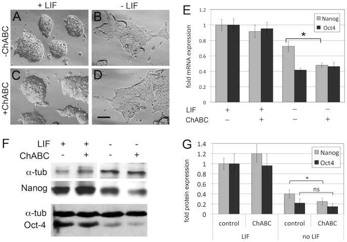
Figure 2. Enzymatic elimination of CS accelerates loss of Nanog expression in differentiating ES cells.
(A–D) Morphology of ES cell cultures in the presence (A, C) or absence (B, D) of LIF, and in the presence (A,B) or absence (C,D) of ChABC. No major morphological changes were observed in ChABC-treated cultures. Scale bar = 20 micrometer. (E) Quantitation of expression of the pluripotency markers Nanog and Oct4 by qPCR. In the presence of LIF, treatment of ES cells with ChABC did not alter expression of Nanog and Oct4. In the absence of LIF, expression of Nanog and Oct4 was downregulated in differentiating ES cells, as expected. Treatment with ChABC lead to a further reduction in Nanog expression levels. (F) Western blot analysis of Nanog and Oct4 protein expression in the presence and absence of LIF and/or ChABC. Alpha-tubulin is shown as loading control. (G) Quantitation of three independent Western blots: ChABC did not affect protein levels in ES cell cultures in the presence of LIF. Withdrawal of LIF caused a reduction of Nanog and Oct4 protein levels, and concomitant treatment with ChABC caused an additional significant reduction in Nanog protein levels, while not significantly affecting Oct4 levels (*p<0.05).

