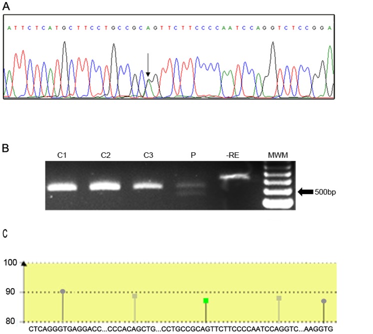
Figure 1. Representative electropherogram and restriction enzyme assay of the novel splice mutation found.
A: Mutation g.1329A>G (c.652-2A>G) changes an acceptor splice site in intron 5. The change A>G is depicted by an arrow. B: BtgI restriction enzyme assay for c.652-2A>G. C: control individuals, P: patient. -RE: control without restriction enzyme. MWM: molecular weight marker. C: Cartoon representation of the bioinformatic results. A fragment of 206 pb ranging from nt 1221 in the 3′ end of exon 5 to nt 1425 in the exon 6 was analyzed using the Human Splicing Finder tool. Only the main sites were included in the diagram. Green square: consensus acceptor site in the intron 5 disrupted by the mutation; grey squares: putative critic acceptor sites located 5′and 3′ of the canonical one; grey circles: consensus donor sites.

