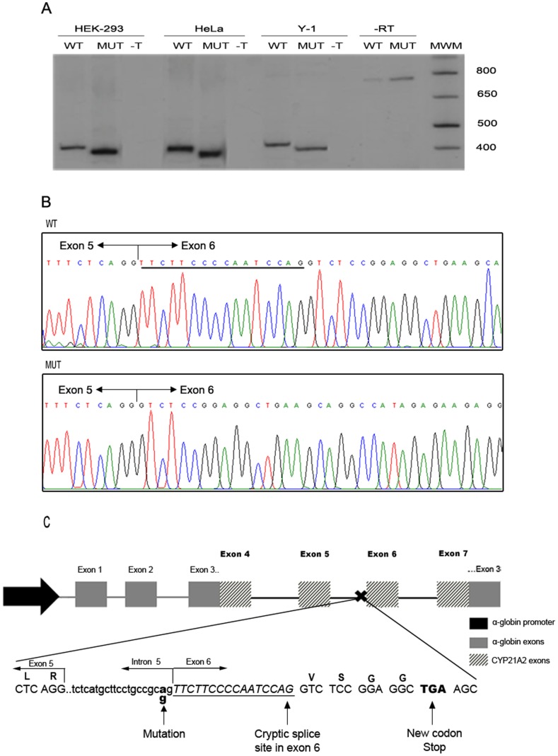
Figure 4. pre-mRNA splicing analysis of wild type (WT) and mutant (MUT) c.652-2A>G CYP21A2 genes.
A: Representative polyacrilamide gel electrophoresis of a Reverse Transcription (RT)- PCR analysis of CYP21A2 minigene mRNAs in 3 cells types: HEK-293, HeLa and Y-1. –T: not transfected. -RT: without reverse transcription: bands observed in both lanes correspond to PCR products from plasmid DNA used as template. MWM: Molecular weight marker. B: Representative electropherograms of the RT-PCR products. Junction of exon 5 and 6 are depicted in each case. Underlined, the 16 nt present in the wild type sequence and absent in the mutant one. C: Diagram summarizing the strategy used and the results obtained. Letters above the sequence represent the codified aminoacids. The 16 nt deletion is in italic and underlined. Dashed arrows represent primers used in the RT-PCR spanning the α-globin and CYP21A2 regions of the minigene.

