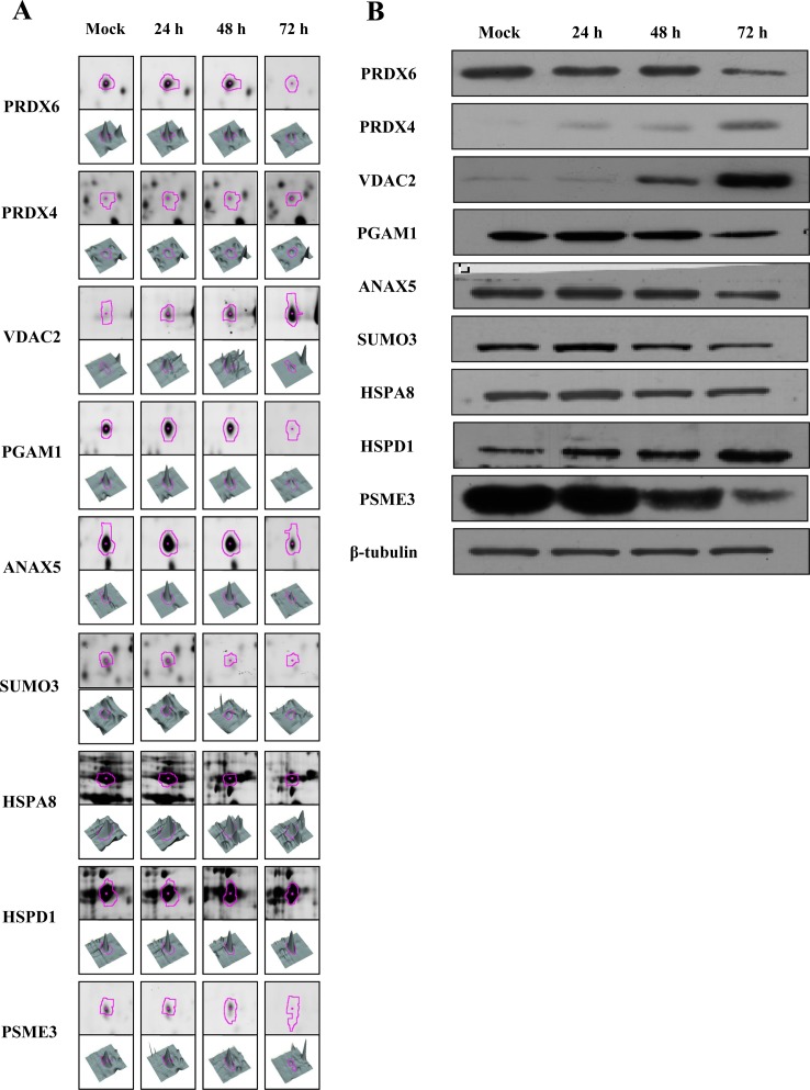
Figure 5. Representative western blot for verification of selected differentially expressed proteins in ARV-infected DF-1 cells.
(A) 2D-DIGE spots (pink outline) and 3D representations of spot intensities for representative upregulated proteins (PRDX4, VDAC2, and HSPD1) and downregulated proteins (PRDX6, PGAM1, ANXA5, SUMO3, HSPA8, and PSME3). (B) Equal amounts of total cell lysates from mock-infected (72 h PI) and ARV-infected DF-1 cells at 24, 48, and 72 h PI were subjected to SDS-PAGE and immunoblot analysis using antibodies against the indicated proteins PRDX6, PRDX4, VDAC2, PGAM1, ANXA5, SUMO3, HSPA8, HSPD1, and PSME3. β-Tubulin was used as an internal control and for normalization.

