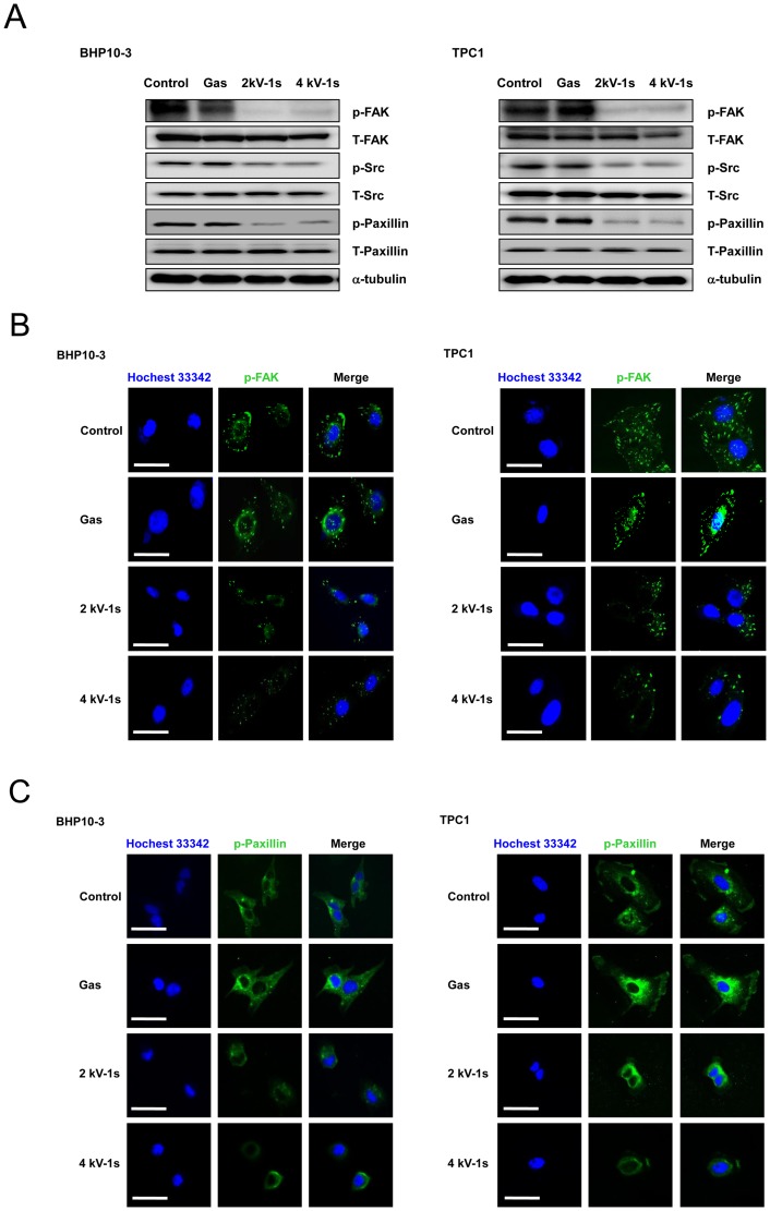
Figure 5. NTP treatment altered the expression of FAK, Src, and paxillin.
(A) Western blotting for p-FAK, p-Src, and p-paxillin. Immunocytochemical assay for (B) p-FAK and (C) p-paxillin. In the control and gas treated-cells, FAK was localized in small adhesion structures at the cell periphery. After NTP treatment, FAK focal accumulation was significantly decreased in both cell lines. Moreover, p-paxillin staining was co-localized with p-FAK staining. NTP treatment also decreased p-paxillin expression. Scale bar = 50 µm. Each figure was representative of three experiments with triplicates.

