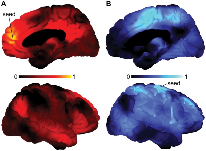
Figure 2. Functional connectivity analysis of the mPFC and MOC cluster.
(A) Figures display right-hemispheric surface mappings of a functional connectivity analysis utilizing the mPFC cluster as seed region. Areas showing increased coupling with the mPFC comprised the PCC, precuneus, MTG and temporal parietal junction. All of these areas correspond to core regions of the DMN. (B) Figures display right-hemispheric surface mappings of a functional connectivity analysis utilizing the MOC cluster as seed region. Areas showing increased coupling with the MOC have been found exclusively in brain regions that spatially correspond to the CEN or SN, while functional coupling with the DMN was absent. The corresponding left-hemispheric mappings are shown in Figure S6. Colorbars represent mean Pearson’s r. Medial prefrontal cortex, mPFC; posterior cingulate cortex, PCC; middle temporal gyrus, MTG; motor cortex, MOC; default-mode network, DMN; central executive network, CEN; salience network, SN.

