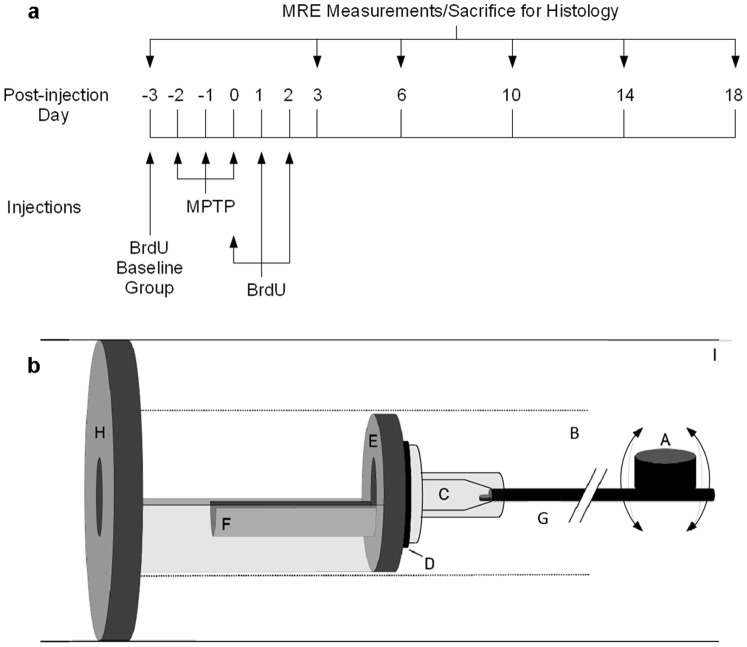
Figure 1. Timeline of the study design and schematic of the mouse MRE apparatus.
The timeline (a) of MPTP and BrdU injections, time-points of MRE measurement and sacrifice for histology. A schematic (b) of the mouse MRE apparatus: (A) driving coil, (B) magnet bore, (C) respiratory mask, (D) rubber bearing, (E) retaining bracket, (F) mouse bed, (G) carbon fiber piston, (H) plastic disk, and (I) Lorentz coil (modified from [16]).

