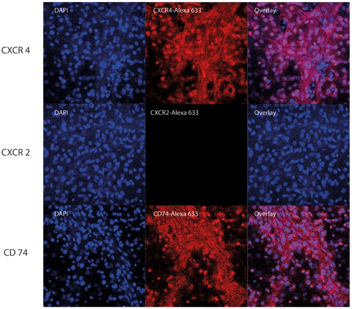
Figure 2. Characterization of MIF receptors by confocal microscopy.
Rat cardiomyocytes were grown in an Ibidi μ-dish for 12 days. They were fixed with paraformaldehyde, permeabilised with Triton X-100 and stained with antibodies against CD74 (A) and CXCR4 (B) and fluorescently labeled secondary antibodies (Alexa 633). Nuclei were stained with Hoechst33342, a cell membrane permeable, DNA-binding fluorophore staining nuclei of cells with blue fluorescence.

