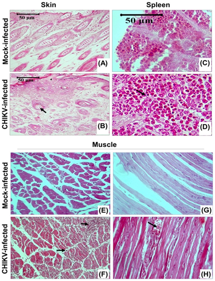
Figure 3. Skin, spleen and muscle pathology of CHIKV infected mice.
Representative picture showing the pathology staining of the tissues (skin, spleen and hind limb muscle) from mock-infected and CHIKV-infected mice on 8th day of post infection. Characteristic histological features are indicated by arrows. Skin of CHIKV-infected mice showed hyperplasia of basal keratinocytes and hyperkeratinisation. Hair follicles showed atrophy with no dividing cells in the matrix. Spleen of CHIKV-infected mice showed considerable lymphoid proliferation and haemorrhage. Muscle sections showed degenerative changes with dark pink stained, atrophied and necrotic muscle fibres, infiltration of neutrophiles and monocytoid cells between the muscle fibres.

