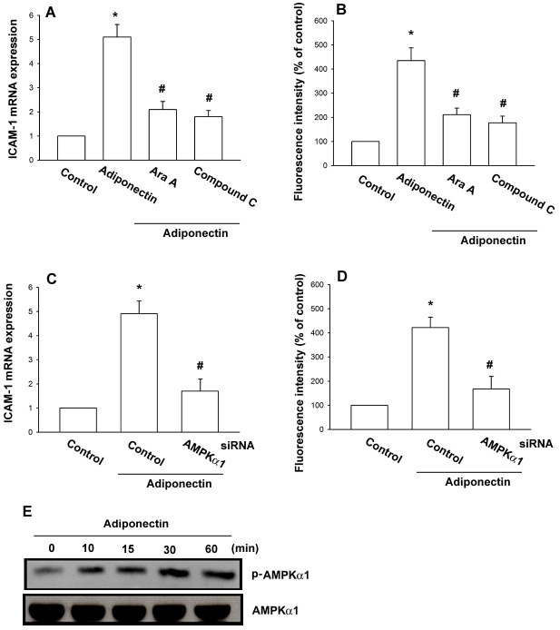
Figure 3. AMPK is involved in adiponectin-induced ICAM-1 expression in synovial fibroblasts.
OASFs were pretreated for 30(0.5 mM) or compound C (10 μM) followed by stimulation with adiponectin (3 μg/ml) for 24 h, and ICAM-1 expression was examined by qPCR (A) and flow cytometry (B). OASFs were transfected with AMPKα1 siRNA for 24 h followed by stimulation with adiponectin (3 μg/ml) for 24 h, and ICAM-1 expression was examined by qPCR (C) and flow cytometry (D). (E) OASFs were incubated with adiponectin (3 μg/ml) for the indicated time intervals and AMPKα1 phosphorylation was determined by western blot. Results are expressed as the mean ± S.E. *p<0.05, compared to basal expression levels. #p<0.05, compared to expression levels in the adiponectin-treated group.

