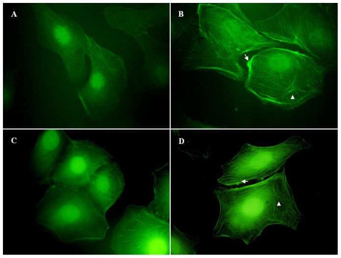
Figure 5. LMW-HA stimulation induces the reorganization of actin filaments in LECs.

SVEC4-10 cells were grown on cover slips and serum-starved overnight. Then the cells were incubated with (B) LMW-HA (3.13 μg/ml) or (A) medium only for 24h. Cells treated with (D) FGF-2 (20 ng/ml) or (C) CSA (3.13 μg/ml) was used as positive or negative control, respectively. After stimulation, cells were fixed on cover slips and stained with FITC-phalloidin. Fluorescence microscope was used to analyze the rearrangement of actin filaments and the changes in cell morphology. All experiments were repeated at least three times and show a representative example. Magnification was ×100. The arrows indicate stress fibers. The arrowheads indicate rosette-like dots.
