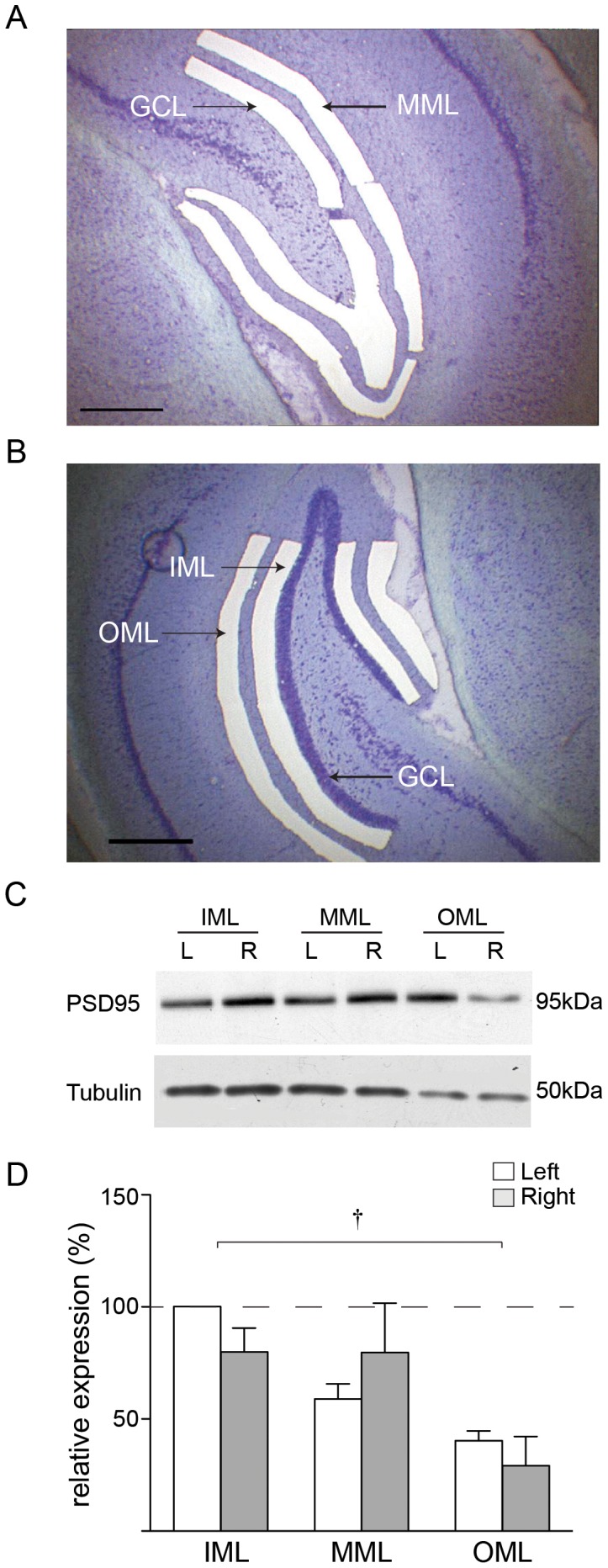
Figure 1. Detection of synaptic proteins in laser microdissected tissue.

(a) Laser microdissection of middle molecular layer (MML) and granule cell layer (GCL) of dentate gyrus from a thionin-stained 35 μm coronal section of rat brain. (b) Laser microdissection of outer (OML) and inner (IML) molecular layers. (c) PSD-95 and tubulin expression in naïve rats: representative Western blot showing PSD-95 and tubulin expression in laser microdissected tissue from one animal. (d) Mean expression of tubulin (n = 4 animals) determined as a percentage of the left IML; † p<0.01 ANOVA.
