
Figure 3. Pathological comparison of tumors from Th-ALKF1174L/Th-MYCN and Th-MYCN mice with abdominal neuroblastoma.
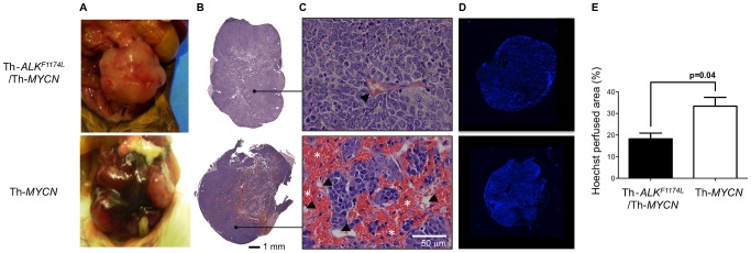
A) Gross pathology, B) composite images, and C) high magnification (x200) images from hematoxylin and eosin stained sections. Note the presence of large hemorrhagic regions filled with aggregated erythrocytes (*, blood lakes) extravasated from blood vessels (arrowed) in the tumor from the Th-MYCN mouse. D) Composite fluorescence images of uptake of the perfusion marker Hoechst 33342 into tumors from Th-ALKF1174L/Th-MYCN and Th-MYCN mice with abdominal neuroblastoma. E) Quantitation of Hoechst 33342 uptake revealed significantly lower functionally perfused vasculature in tumors of Th-ALKF1174L/Th-MYCN mice (n = 5) compared with tumors in Th-MYCN mice (n = 5). Data are mean ±1 s.e.m, p, Student's 2-tailed unpaired t-test with a 5% level of significance.