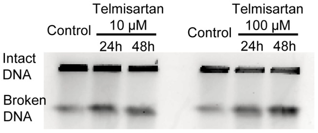
Figure 5. Analysis of telmisartan-induced DSB formation in HHUA cells.

DSB formation was analyzed by PFGE. HHUA cells collected into agarose plugs, and their DNA was separated by size on an agarose gel. Under the electrophoresis conditions used, high-molecular-weight genomic DNA remained in the well, whereas lower-molecular-weight DNA fragments (several Mbp to 500 kbp) migrated into the gel and were compacted into a signal band.
