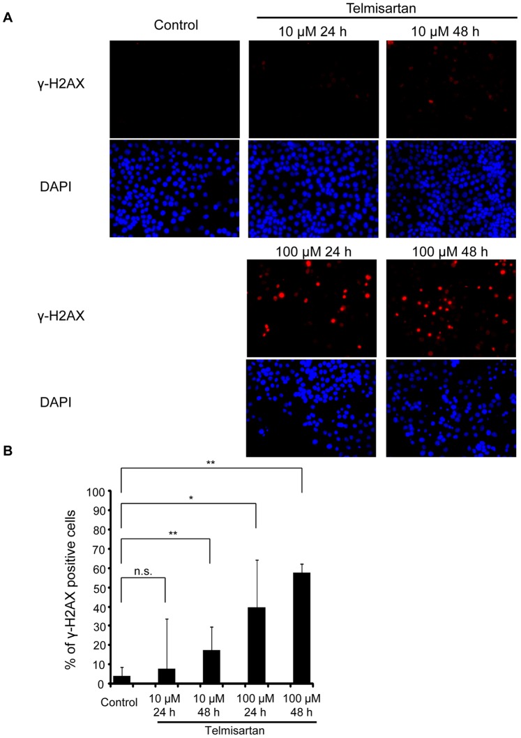
Figure 6. Immunofluorescent staining of γ-H2AX.
(A) Time and dose dependency of histone H2AX phosphorylation by telmisartan. HHUA cells were treated with 10 or 100 μM telmisartan for indicated times followed by immunostaining using an anti-γ-H2AX antibody. Their nuclei were revealed by DAPI staining. (B) Percentage of γ-H2AX-positive cells. HHUA cells were treated with 10 or 100 μM telmisartan for 24 h or 48 h. Control cells were treated with vehicle alone. The telmisartan-treated cells had significantly higher levels of fluorescence compared to the untreated cells. Results = means ± SD of three independent experiments. Columns, means; bars, SDs. *P<0.05 vs. control, **P<0.01 vs. control.

