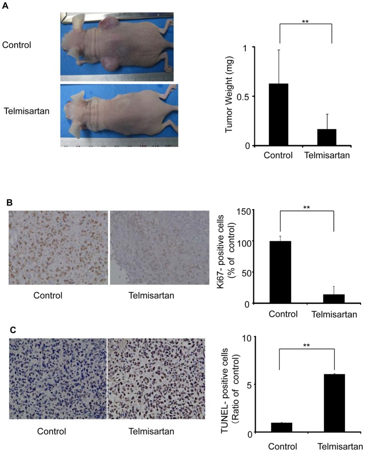
Figure 7. HHUA tumors in nude mice treated with telmisartan.
(A) HHUA cells (5×10⁶) were bilaterally subcutaneously injected into the trunks of nude mice, forming two tumors per mouse. The mice were divided randomly into control and experimental groups. Telmisartan (100 μg/mouse) or diluent (control) was administered intraperitoneally for 5 days a week for 7 weeks. After 7 weeks of therapy, the tumors were removed from each mouse and weighed. The tumor weights in the two groups were significantly different. Columns, means; bars, SDs. **P<0.01 vs. control. (B) The effect of telmisartan on HHUA tumors in nude mice. The endometrial carcinoma cells from mice treated with telmisartan showed significantly weaker staining for Ki-67 compared to the control endometrial carcinoma cells. (C) Endometrial carcinoma cells from the mice treated with telmisartan underwent apoptosis (TUNEL-positive). All assays were performed three times. Columns, means; bars, SDs. **P<0.01 vs. control.

