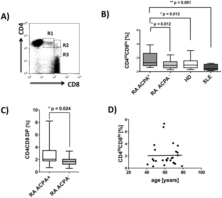
Figure 1. Increased frequencies of peripheral CD4CD8 DP T cells in ACPA+ rheumatoid arthritis patients.
PBMC were isolated from RA patients (ACPA+, n = 37, ACPA−, n = 20), healthy donors (n = 36), and patients with SLE (n = 8), and FACS analyses from live cells (PI staining used for exclusion of dead cells) were performed. Analyses for CD4CD8 double positive T cells are always pregated on CD3 positive T cells. A) Representative FACS plot from one RA patient, showing CD4CD8 double positive T cells, R1 = CD4hiCD8lo, R2 = CD4hiCD8hi and R3 = CD4l°CD8hi B) Comparison of frequencies of CD4hiCD8loT cells in PBMCs of ACPA+/− RA, HD and SLE patients. C) Total CD4CD8 DP T cells in ACPA+ (n = 19) and ACPA− (n = 20) RA patients. D) Correlation analysis for frequencies of CD4hiCD8lo T cells with age in ACPA+ RA patients. Box plots depict median, interquartile range and 10–90 percentile. Significance as given, *p<0.05, **p<0.01.

