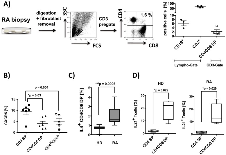
Figure 4. CD4CD8 double positive T cells are present in the synovium of RA patients and produce T helper 2 like cytokines.
A) Single cell suspensions were prepared from synovial biopsies from RA patients (n = 4) and analyzed by flow cytometry using anti-CD19, anti-CD3, anti-CD4 and anti-CD8 monoclonal antibodies (PI staining used for exclusion of dead cells). Representative FACS plots are depicted. Each data point represents one experiment. B) PBMCs from RA patients (n = 6) were isolated and FACS analysis from live cells were performed (PI staining used for exclusion of dead cells). Cells were pre-gated on CD3+ T cells, and CXCR5 expression was analyzed in the subpopulation indicated. C) PBMC were stimulated in vitro for 4 hrs with Cytostim. Subsequently, an IL-4 specific secretion assay was performed, and expression of CD4 and CD8 was determined in vital cells (PI staining used for exclusion of dead cells) pre-gated on CD3. Percentage of IL-4 producing CD4CD8 double positive T cells in HD (n = 7) and RA patients (n = 8) is given. D) PBMCs from HD (n = 4) and RA (n = 4) were stimulated with PMA/Iono for 4 hrs, and intracellular staining for IL-21 was performed. Cells are counterstained with anti-CD3, anti-CD4 and anti-CD8. Depicted is the percentage of cytokine producers in total CD4CD8 DP T cells and in CD4 SP cells. Box plots in C) and D) show median, interquartile range, and 10–90 percentiles. Significance as given, *p<0.05.

