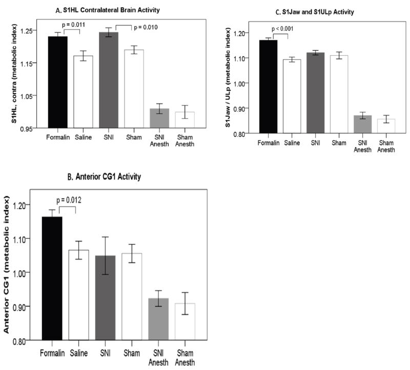
Figure 6.
Anatomically defined regions of interest (ROIs) for formalin, spared nerve injury (SNI), SNI anesthetized, and respective controls. (A) primary somatosensory (S1) hindlimb contralateral brain activity differences are seen in both formalin and SNI, but not SNI anesthetized versus controls. (B) Anterior cingulate area 1 (Cg1) difference is seen in formalin vs. controls, but not in either SNI group versus controls. (C) S1 jaw & S1 upper lip difference is seen in formalin versus controls, but not in either SNI group versus controls. Error bars +/− 1 S.E.

