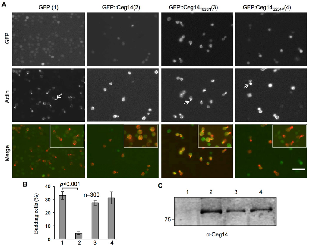
Fig. 4. Ceg14 interferes with daughter cell budding in yeast.
A. Yeast strains harboring GFP, GFP-Ceg14, GFP-Ceg14T623N, or GFP-Ceg14G234V under the control of the Pgal promoter were induced by galactose for 6 hrs. Fixed, permeabilized cells were stained with Texas Red-X phalloidin prior to image acquisition and scoring of budding cells. Note that in cells expressing GFP or GFP fusions of the mutants, the actin staining signals mostly localized to the growing tips of the daughter cells (arrows in the middle panel of the first, third and fourth column of images), whereas in cells expressing GFP-Ceg14, actin was lining along the plasma membrane (arrows in the middle of the second column of images). Bar, 10 µm. B. The number of budding cells was quantitated under a microscope in stained samples done in triplicate, 300 cells were scored in each sample. Similar results were obtained in multiple independent experiments and data shown were from a representative experiment. C. Expression of the fusion proteins in the samples. Total protein from a portion of the cells used for immunostaining resolved by SDS-PAGE was probed with a Ceg14 specific antibody.

