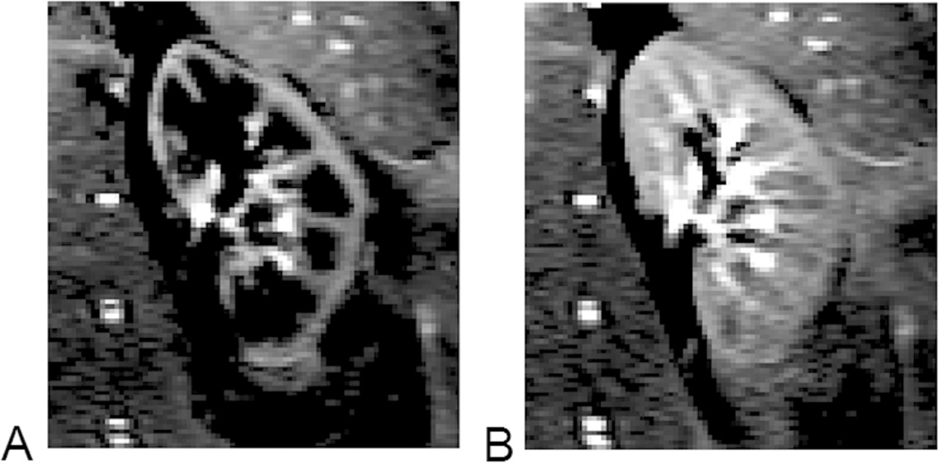
Figure 3.
Difference images obtained from renal ASL scans. A) Acquired at 800 ms after arterial blood labeling, when the labeled blood is mostly in renal cortex; B) 1000 ms after the labeling, and some labeled blood reaches renal medulla. The images were acquired by a modified TrueFISP FAIR ASL sequence (8 averages, acquisition time ~24 sec, with breath hold).

