Figure 5.
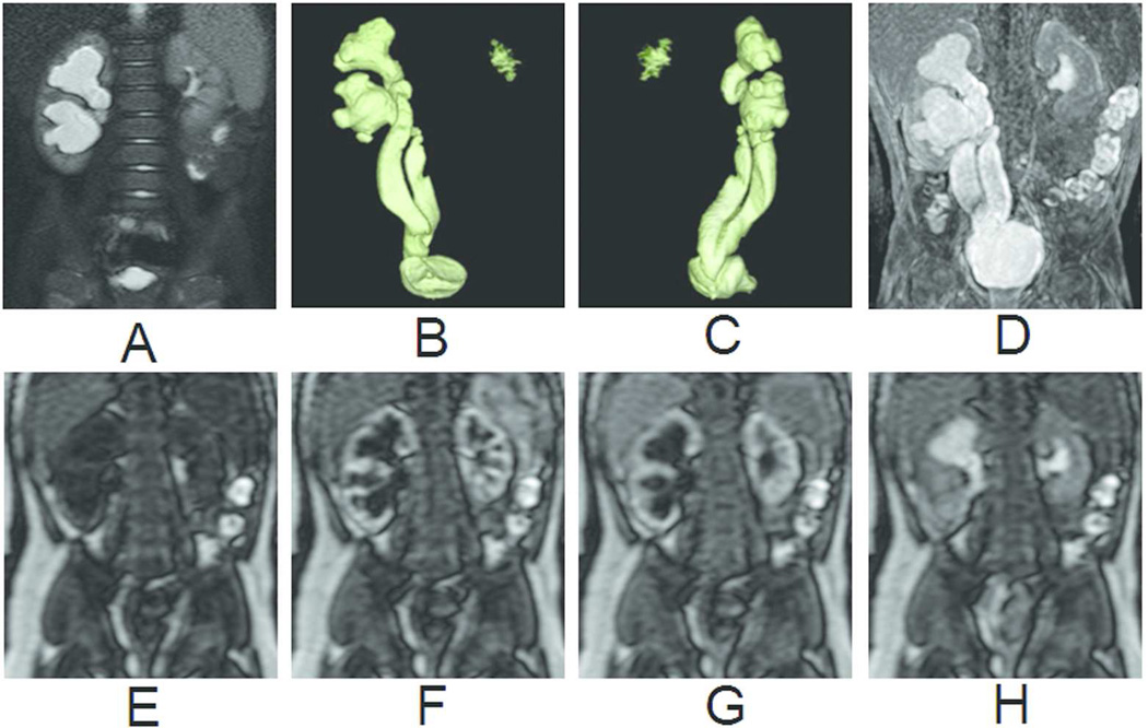
Primitive right megaureter on a bifid ureter in a 6-month old boy. (A) T2-weighted image with fat saturation. (B) Coronal view of volume-rendered T2-weighted images. (C) Oblique view of volume-rendered T2-weighted images. (D) Maximum intensity projection of T1-weighted images at excretory phase. (E) Renography before contrast arrival. (F) Renography at arterial phase. (G) Renography at tubular phase. (H) Renography at excretory phase. Symmetric enhancement and excretion of contrast bilaterally suggests that the marked dilatation of the right collecting system and ureter is not a functional obstruction.

