
Figure 3.
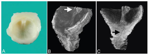
Type III posterior urethral valve (urethral diaphragm) imaged by optical projection tomography (case 3). A. Gross specimen consisting of transected posterior urethra, portion of verumontanum, and inferior urethral crests extending to obstructive diaphragm. B. Superior-oblique view of posterior urethra showing inferior portion of verumontanum (arrow) and inferior urethral crests that lead to obstructive diaphragm. C. Coronal section of urethra showing same structures as A plus stenotic, discontinuous urethra (arrow). A color version of this figure is available online.