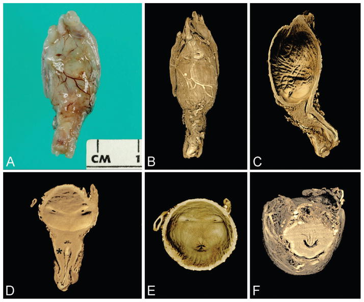
Figure 4.
Control specimen imaged by microCT (23 weeks). A. External view of anterior bladder with umbilical arteries present laterally and proximal penile shaft inferiorly (gross specimen). B. Rendered external view (same orientation as A). C. Virtual section of bladder and urethra in median plane. D. Virtual section of bladder and urethra in coronal plane; note insertion of ureters and verumontanum (asterisk) with inferior urethral crest. E. Base of urinary bladder showing urethral orifice centrally and 2 ureteral orifices laterally. F. Virtual section of posterior urethra in transverse plane showing verumontanum, ejaculatory ducts, and prostatic utricle (U-shaped urethral lumen is normal configuration at this level). A color version of this figure is available online. Color and shadowing are computer-generated.

