
Figure 7.
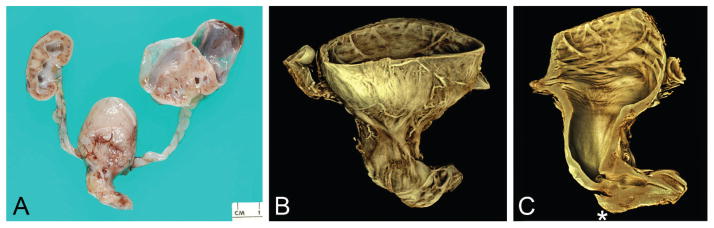
Atypical kinking of posterior urethra with poststenotic dilatation and more distal obstructing diaphragm (case 10). A. Gross specimen showing hydronephrosis, with large perirenal cyst (urinoma), hydroureter, megacystis, dilated posterior urethra, and proximal penis. B. Rendered volume after microCT imaging of sectioned bladder, posterior urethra, and proximal penis. C. Virtual section in median plane showing dilated urinary bladder and posterior urethra; from proximal to distal, note regions of urethral stenosis followed by poststenotic dilatation, obstructing diaphragm, and patent urethra. (This distal diaphragm is consistent with a “congenital obstructive posterior urethral membrane,” or COPUM, as described by others [20].) When scanned, the lumen of the bladder/proximal urethra and lumen of the more distal urethra did not lie in the same plane; this was overcome by manually defining separate planes for the tissue with the software, the joining of which resulted in the faint oblique line visible across the stenotic urethral lumen (asterisk). A color version of this figure is available online. Color and shadowing are computer-generated.