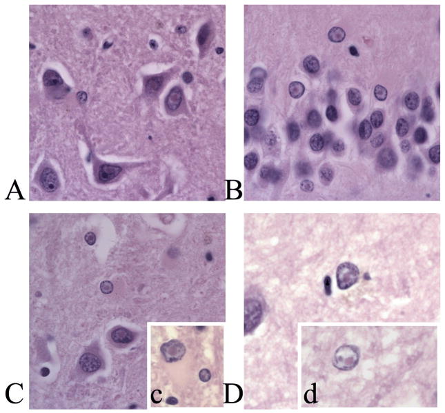
Figure 1.
A. Intranuclear inclusions in pyramidal cells in CA3b of Case 2. B. Intranuclear inclusions in granule cells of the medial blade of the dentate gyrus of Case 2. C. Intranuclear infusions in pyramidal cells and neurons in the hilus of case 6. c. Inset, intranuclear inclusion in neuron and astrocyte in the white matter adjacent to layer VI of the frontal cortex of Case 6. D. Intranuclear inclusion in an astrocye in layer II if the frontal cortex of Case 1. d. Inset, granule cell from layer IV of the frontal cortex in Case 1. All plates are at 1000X magnification. Scale bar = 50μm.

