
Figure 6.
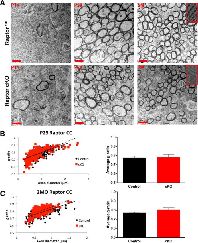
In contrast to spinal cord, in Raptor cKO corpus callosum, myelin sheath compaction was relatively normal. A, Electron micrographs in the corpus callosum of floxed Raptor (black) and Raptor cKO (red) mice at P14 (initiation of myelination), P29 (peak of active myelination), and 2 month (2M; myelin maintenance phase). B, Quantification of the g-ratio as a scatter plot with axonal diameter on the x-axis and the g-ratio on the y-axis for the P29 and 2 month floxed Raptor and Raptor cKO animals (B, C). At P14, there were few myelinated axons (large caliber) in either group. Note high-magnification images of myelin at P29, where there were subtle deficits in myelin compaction in the Raptor cKO mice, but there is not an overall reduction in myelin sheath thickness. At 2 months (2MO), the axonal caliber and myelin thickness was comparable between Raptorfl/fl and Raptor cKO mice (C). Scale bars: A, low-magnification images, 1 μm; high-magnification image insets, 100 nm. Values displayed as ±SEM (n = 3, *p < 0.05, **p < 0.005, ***p < 0.001).