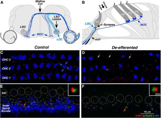
Figure 2.
Immunostaining of the cochlear efferent and afferent innervation in normal and de-efferented cases. A, Brainstem schematic shows the central origins of LOC and MOC efferents. Efferent pathways were lesioned via midline cut of MOC fibers at the floor of the IVth ventricle or by neurotoxin injection into the LSO where LOC fibers originate. B, Organ of Corti schematic shows the peripheral targets of cochlear efferents. MOC terminals synapse with outer hair cells (OHCs); LOC terminals synapse with auditory nerve fibers (ANFs) near their afferent synapses with the inner hair cell (IHC). Each IHC–ANF synapse is identifiable by its presynaptic ribbon (red) and postsynaptic glutamate receptors (green). The small population of cochlear nerve fibers (<5%) that contact OHCs are not shown. C–F, The success of de-efferentation was assessed by immunostaining cochlear efferent terminals with antibodies for VAT (blue); IHC-ANF synapses were stained for a protein in the presynaptic ribbon (CtBP2; red) and an AMPA postsynaptic glutamate receptor (GluA2; green). Each image is a maximum projection of a focal series through the synaptic region of the OHCs (C, D) or IHCs (E, F), from the viewing angle schematized in B. Each control OHC (C) is innervated by a cluster of 1–4 MOC terminals (e.g., blue arrows); in the de-efferented ear (D), many OHCs have no MOC endings, but presynaptic ribbons remain near the ANF terminals (e.g., at arrows). In the IHC area (E, F), individual sensory cells are difficult to distinguish, so their nuclei are indicated by dashed circles. In the control ear (E), cochlear nerve synapses are seen as paired red-green puncta in the subnuclear zone, just above the LOC terminals in the inner spiral bundle. In the de-efferented ear (F), synaptic counts are reduced, and the LOC innervation is almost completely eliminated. In each IHC panel, one synapse (at the arrow) is shown at higher magnification in the inset. Scale bar in F applies to all large images. Scale bar in one inset applies to both. All images are from the middle of the cochlear spiral (16 kHz). D is from a midline-cut case; F is from an LSO lesion case.

