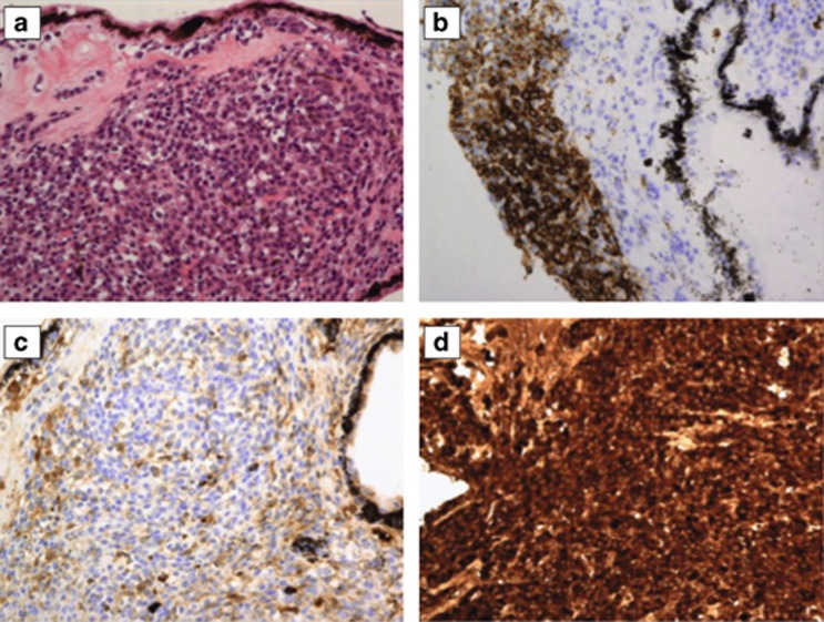
Figure 2.
Histopathological examination was performed on a trans-scleral incisional biopsy specimen of the ciliary body tumour. (a) Hematoxylin–eosin-stained section showing a diffuse infiltrate of small plasma cells and lymphocytes. (b) Immunohistochemical stain for CD20 showing that the infiltrate consists predominantly of B cells. (c, d) Immunohistochemical stain for immunoglobulin light chains showing monotypical expression for kappa; monoclonality was confirmed using IgKappa-PCR. Taken together these features are consistent with extranodal marginal zone B-cell lymphoma of the ciliary body.

