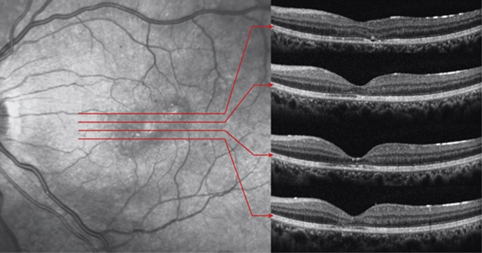
Figure 2.
Infrared reflectance (IR) image and corresponding optical coherence tomography (OCT) images of a contusion injury and subsequent commotion retinae (Case 14). The IR image showed diffuse macular hypo-reflectance and focal hyper-reflective lesions therein. The OCT images revealed the following: (1) the cone outer segment tip (COST) defects corresponded to hypo-reflective areas on the IR image and (2) the reflectivity loss of multi-layers in the COST, inner and outer segment junction, and external limiting membrane corresponded to hyper-reflective lesions on IR image.

