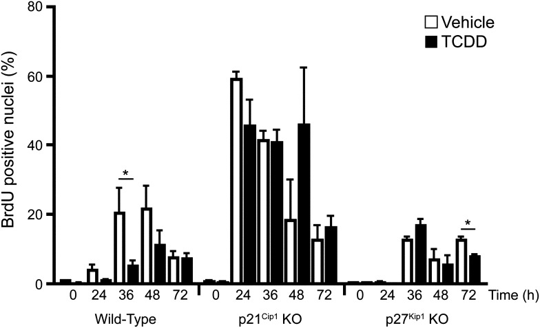
Fig. 1.
Analysis of hepatocyte proliferation during liver regeneration in vehicle or TCDD-treated wild-type, p27Kip1 KO mice, or p21Cip1 KO mice. Mice were treated for 24 hours with vehicle or TCDD prior to PH (t = 0 hours). Animals were pulsed with BrdU 2 hours before being euthanized at indicated time points. BrdU-positive nuclei were counted in six random immunohistochemical fields (>300 cells/field) in liver tissue from each mouse. Data represent the average percent positive nuclei from three animals per treatment group for each genotype, and are representative of three separate experiments. Data are plotted as the mean ± S.E.M. *Significant difference between the vehicle and TCDD-treated groups (P < 0.05).

