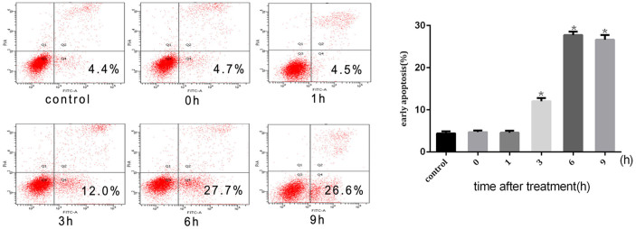
Figure 2. Intense heat stress-induced apoptosis.

HUVEC cells were treated with 43°C for 2 h, culture media were replaced with fresh media and the cells were further incubated for different times as indicated, and apoptosis was analyzed by flow cytometry using Annexin V-FITC/PI staining. Data are presented as means ± SD of three separate experiments. The asterisk indicates a significant difference between control(37°C) and test groups(43°C), *p < 0.05.
