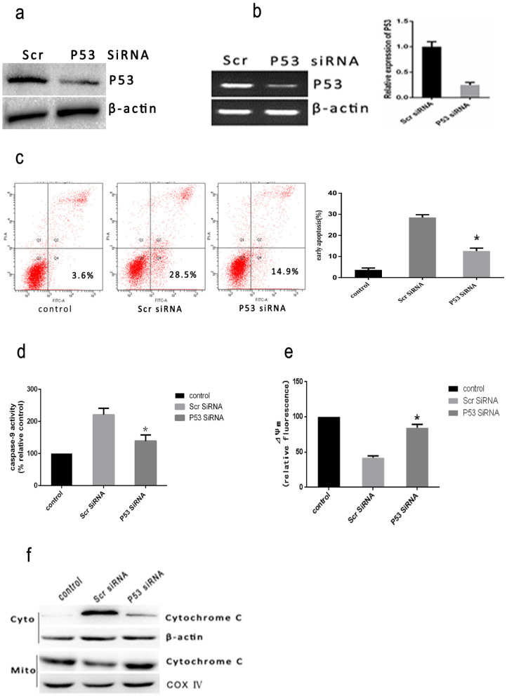
Figure 5. Heat stress-induced apoptosis in P53 siRNA transfectant HUVEC cells.
(a) HUVEC cells were transfected with scrambled siRNA (Scr) or P53 siRNA (P53). Western Blots of P53 protein expressed(cropped) in HUVEC transfectant cells.β-actin was run as an internal control. (b) RT-PCR analysis of P53 in HUVEC transfectant cells. Total RNA was isolated from HUVEC transfectant cells and P53 mRNA levels were determined by RT-PCR and analyzed on agarose gel electrophoresis. β-actin from the same samples was amplified as control. (c–f) HUVEC transfectant cells were treated with 43°C for 2 h with the cells further incubated for 6 h, and (c) apoptosis was analyzed by flow cytometry using Annexin V-FITC/PI staining. (d) Enzymatic activity of caspase-9 was measured in cell lysates using fluorogenic substrate Ac-LEHD-AFC and was expressed relative to the control at 37°C (100%).(e) The loss of ΔΨm was measured by JC-1 and flow cytometry. (f) Intracellular location of cytochrome C was determined by Western Blots(cropped). COX IV, mitochondrial loading control. Each value is the mean ± SD of three separate determinations. *P < 0.05 versus scrambled siRNA-transfected cells exposed to heat stress.

