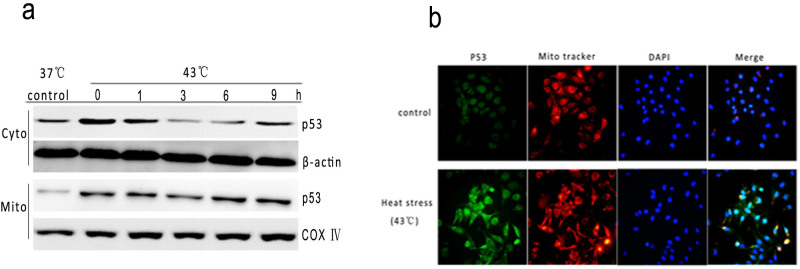
Figure 6. Intense heat stress induces translocation of p53 from nucleus to mitochondria.

(a) To determine localization of p53, HUVEC cells were treated with 43°C for 2 h, further incubated for different time as indicated, and the levels of p53 (mitochondrial and cytosolic fraction) proteins were assessed by Western blot using corresponding antibodies(cropped). COX IV, mitochondrial loading control. (b) immunofluorescence localization of p53 to mitochondria in heat-treated cells. HUVEC cells were treated with 43°C for 2 h, and stained with anti-p53 antibody (green), MitoTracker (red) and nuclear(blue). Merged images are shown in the right panels.
