
Figure 8.
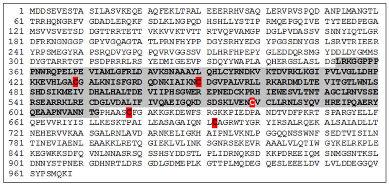
Mapping of S-nitrosation sites in p120. Protein sequence from human p120. Purified p120 was S-nitrosated with GSNO for 30 minutes, subjected to biotin switch assay followed by in-solution trypsin digest. The region that potentially interacts with VE-cadherin is shown in gray background. Cysteine 579 (white letter, red background) was 100% S-nitrosated according to mass spectrometry. Other cysteines (red background) were not S-nitrosated in 100% of the assays. Unlabeled cysteines were not S-nitrosated. (See Supplemental Data for experimental details).