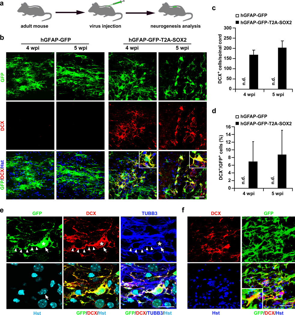
Figure 1. Induction of DCX+ cells in the adult mouse spinal cord.
(a) Experimental scheme. (b) DCX+ cells are detected in animals injected with lentivirus expressing SOX2 but not control GFP at 4 or 5 weeks post viral injection (wpi). Nuclei were counterstained with Hoechst 33342 (Hst) (c–d) Quantification of SOX2-induced DCX+ cells around the virus-injected regions in adult spinal cords (mean+s.d.; n=3 mice per group; n.d., not detected). (e) Confocal images of a representative DCX+ cell (indicated by arrows). SOX2-induced DCX+ cells are traced by the co-expressed GFP, indicating an origin of virus-infected cells. They are also co-labeled by TUBB3 and have bipolar or multipolar processes. Asterisks and arrowheads indicate cell bodies and neuronal processes, respectively. (f) Representative images of DCX+ cells induced in the spinal cord of aged mice (>12 months). Scales: 50 µm (b, f) and 20 µm (e).

