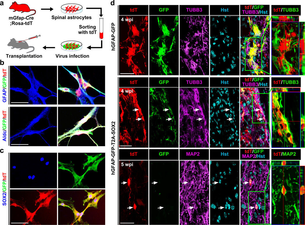
Figure 4. In vivo conversion of engrafted astrocytes to neurons.
(a) Experimental scheme. Spinal astrocytes cultured from mGfap-Cre;Rosa-tdT mice were purified based on tdT expression. Three days post viral infection, cells were transplanted into the spinal cord of NSG mouse. (b) Immunocytochemistry showing the expression of GFAP and Aldolase c (Aldc), markers for astrocytes, in purified tdT+ cells. (c) Immunocytochemistry confirming SOX2 expression in spinal astrocytes infected with the hGFAP-GFP-T2A-SOX2 virus. (d) Confocal images showing astrocyte-derived neurons in the spinal cord transplanted with the SOX2 but not control virus-infected astrocytes. Orthogonal views of cells in the boxed regions are shown in the right panels. Astrocyte-derived neurons (tdT+TUBB3+ or tdT+MAP2+) are indicated by arrows. Scales: 60 µm (b,c) and 30 µm (d).

