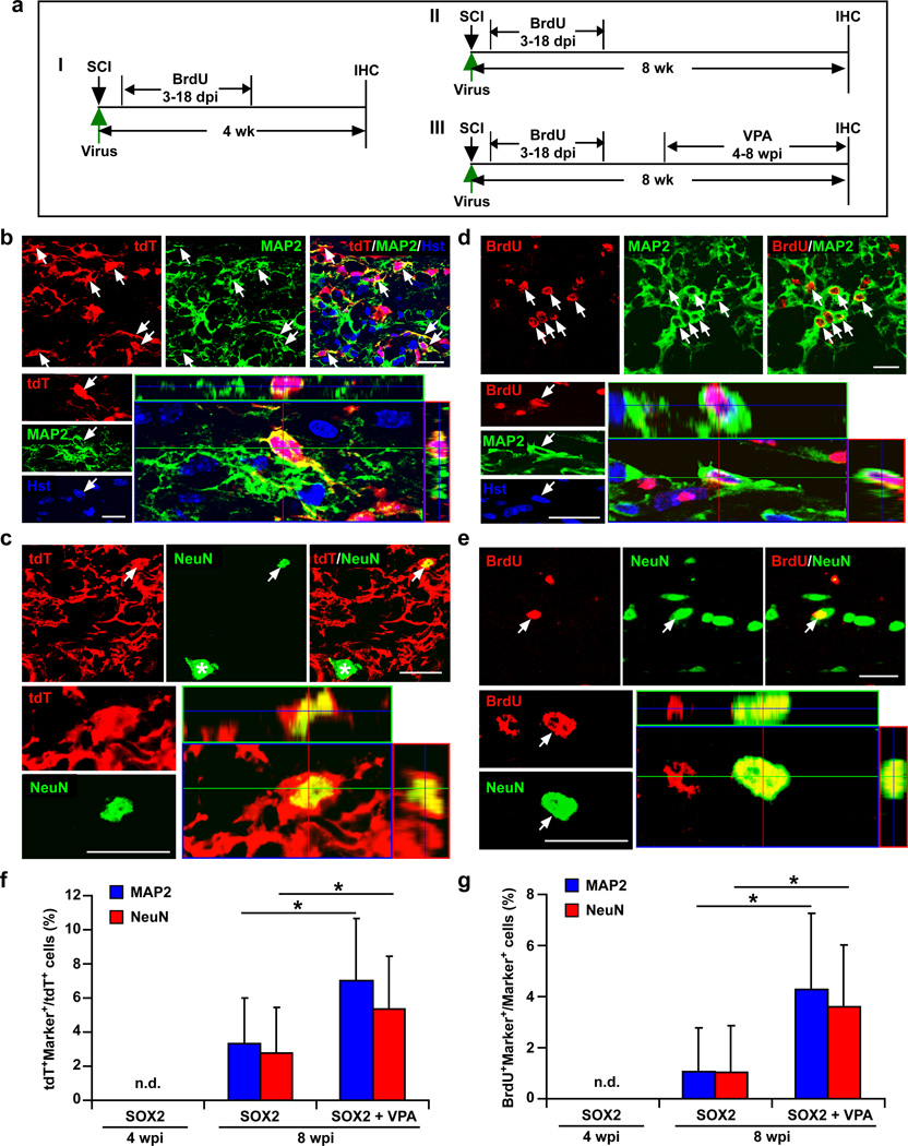
Figure 8. Maturation of SOX2-induced new neurons in the injured adult spinal cord.
(a) Experimental scheme. mGfap-Cre;Rosa-tdT mice were injected with hGFAP-SOX2 or a control virus immediately after injury and analyzed by IHC at 4 (I) or 8 (II, III) wpi (dpi, days post viral injection; VPA, valproic acid). (b–c) Expression of the mature neuronal markers MAP2 (b) or NeuN (c) in tdT+ cells in SOX2 virus-injected spinal cords at 8 wpi. tdT-traced MAP2+ or NeuN+ cells were not detectable in control virus-injected spinal cords. Orthogonal views of cells with expression of the indicated markers are also shown. Compared to endogenous spinal motoneurons (indicated by an asterisk in c), the SOX2-induced neurons are interneuron-like with a smaller soma (indicated by an arrow in c). (d–e) SOX2-induced mature neurons pass through a proliferative stage. Mice were treated with BrdU at 3–18 dpi and analyzed at 8 wpi. Orthogonal views of BrdU-traced mature neurons are also shown. (f–g) Quantification of SOX2-induced mature neurons in the injured adult spinal cord (mean+s.d.; n=5 mice per group; *p<0.01 by Student’s t-test). Scales: 20 µm (b–e).

