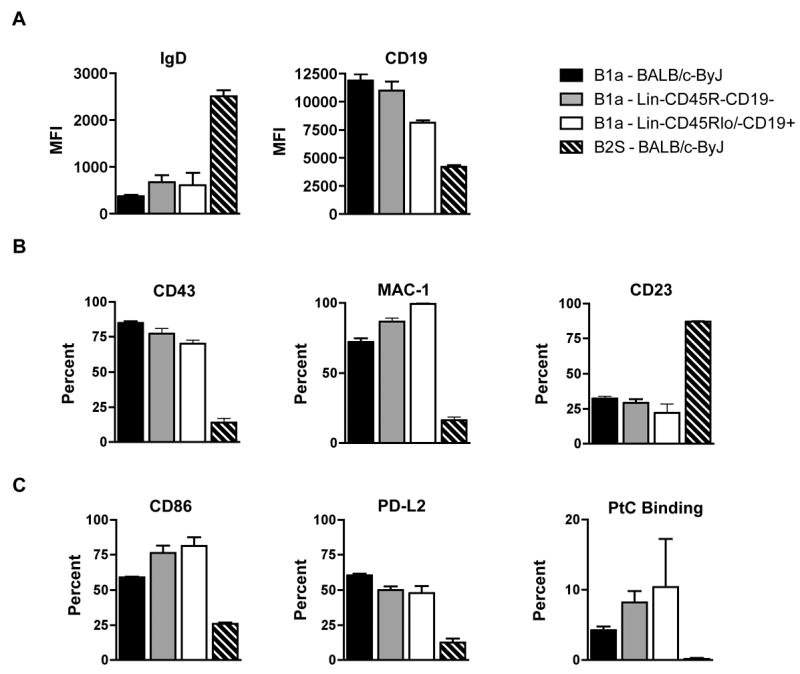
Figure 2. Peritoneal B cells derived from Lin-CD45R-CD19- fetal liver cells phenotype as B-1a cells.

Peritoneal IgMa+CD45RloCD5+ cells derived from Lin-CD45R-CD19- fetal liver cells were analyzed for surface markers typically found on peritoneal B-1a cells. These cells were compared to peritoneal B-1a and splenic B-2 cells from 3-month old BALB/c-ByJ mice in addition to peritoneal IgMa+CD45RloCD5+ cells derived from Lin-CD45Rlo/-CD19+ fetal liver cells. (A) MFI values of IgD and CD19 are shown. (B) Percent of peritoneal IgMa+CD45RloCD5+ cells or splenic IgMa+CD45RhighCD5- cells expressing CD43, Mac-1 (CD11b), or CD23 are shown. (C) Percent of peritoneal IgMa+CD45RloCD5+ cells or splenic IgMa+CD45RhighCD5- cells expressing CD86, PD-L2, or PtC liposome binding are shown. The data shown are an average of six independent experiments with 3-5 mice per group in each experiment.
